Cell diagrams for medical teaching
Labeled cell and tissue figures for education — generate an editable draft from a plain-text description.
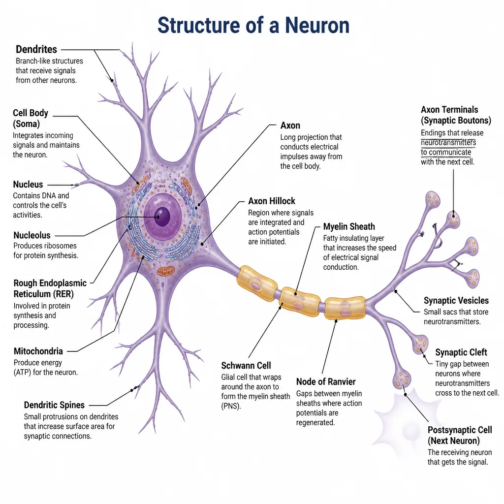
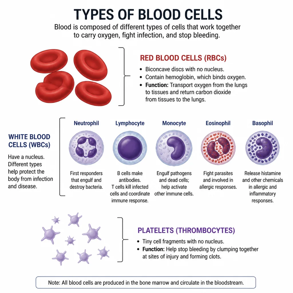
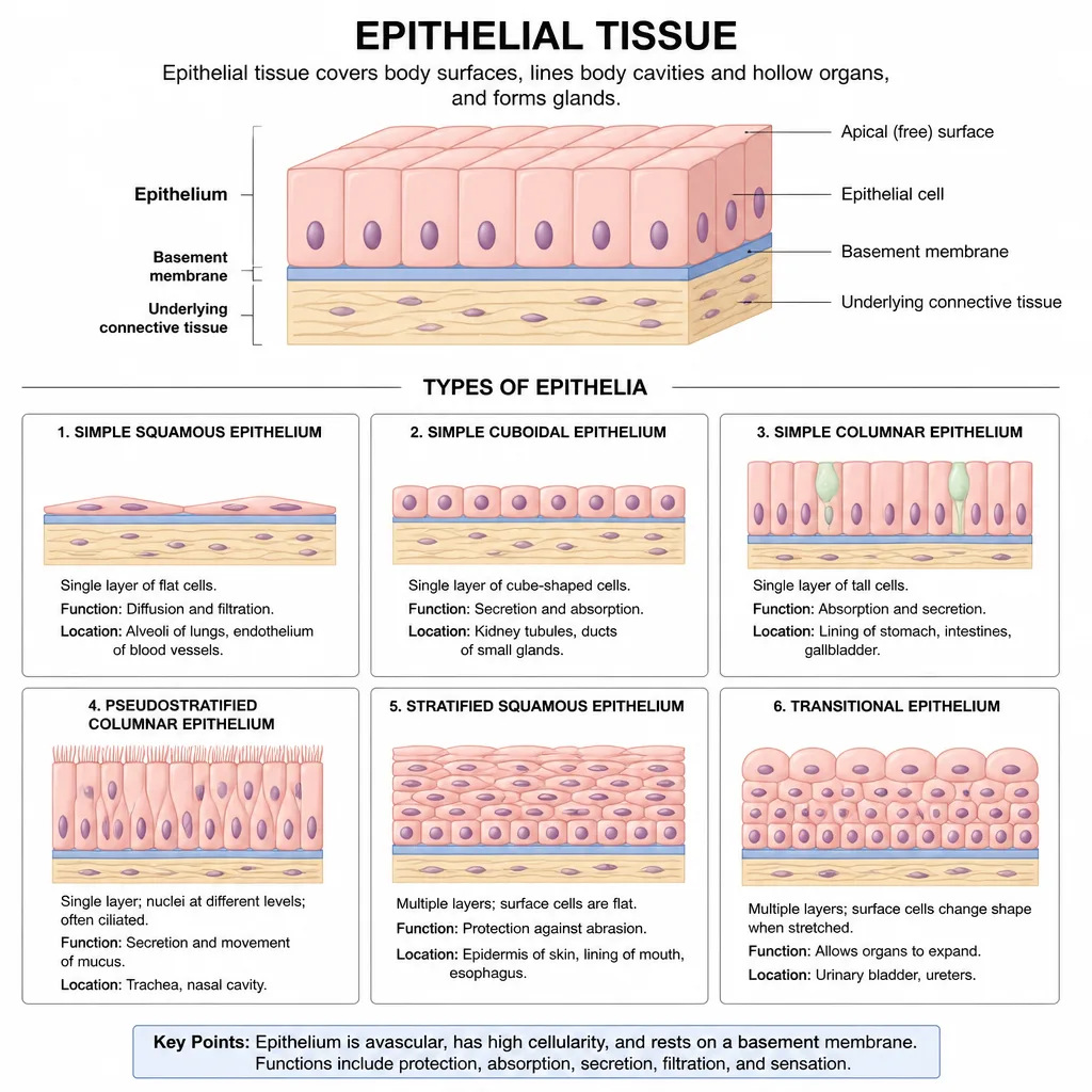
This gallery collects cell and tissue diagram examples from MedicalFigs — neurons, blood cell types, and epithelial tissue among them — the kind of medical cell diagram you need for a histology lecture, a study guide, or a patient explanation of what is happening at the microscopic level. Browse the examples, then describe the cell or tissue you want and generate an editable labeled draft.
Cellular subjects are where a good diagram earns its keep, because the real thing is invisible to the naked eye and every textbook draws it a little differently. MedicalFigs lets you set the terms: name the cell, the structures you want labeled, and the audience, and you get a clean first draft to adjust. Try "a labeled neuron showing dendrites, axon, and myelin sheath for a first-year class" and pare it back to whatever your slide needs.
These cell diagrams support education and communication only. They are stylized teaching figures, not micrographs or validated scientific references, so confirm the details against your course material before teaching from them. When you need the bigger picture, step up to the organs or human anatomy galleries, or return to the medical library to browse every category.
MedicalFigs is for education and communication only. It is not a medical device and does not process patient health information. Generated diagrams are not validated clinical references — review every figure before use.
Can I make custom cell diagrams?
Yes. Describe the cell type and labels; MedicalFigs generates an editable draft.
Are these research-grade molecular figures?
MedicalFigs focuses on clinical teaching and communication. For molecular icon libraries, compare workflows on our BioRender pages.
Can I use these in nursing education?
Yes, for education and communication after your accuracy review.
Where is the full library?
The medical illustration library links all category galleries including cells.
Are these drawn from real micrographs?
No. They are stylized, labeled teaching illustrations generated from your description, not photomicrographs. Use them to explain structure and concepts, and pair them with real imagery when accuracy to a specimen matters.